Anatomy Of Ribs Posterior - Ribs Radiology Reference Article Radiopaedia Org / The ribs are elastic arches of bone, which form a large part of the thoracic skeleton.
byAdmin•
0
Anatomy Of Ribs Posterior - Ribs Radiology Reference Article Radiopaedia Org / The ribs are elastic arches of bone, which form a large part of the thoracic skeleton.. The ribs are elastic arches of bone, which form a large part of the thoracic skeleton. Each rib articulates posteriorly with two thoracic vertebrae by the costovertebral joint. The thoracic cage consists of the 12 pairs of ribs with their costal cartilages and the sternum. The posterior cecal artery is located in the abdomen near the lower intestines. Its anterior ramus forms the intercostal nerves.
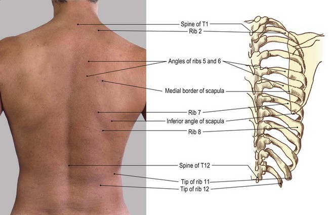
Test your knowledge about the ribs anatomy here It is important to review the anatomy of the chest wall and thoracic cavity, as you will use anatomic landmarks to document the location of respiratory assessment findings. It is split into superior and inferior fibres. Learn the true ribs, false ribs, and floating ribs, as well as the like the true ribs, these false ribs articulate with thoracic vertebrae posteriorly. Illustrations in anterior and posterior view of male torso and back, allowing the lines and regions used in surface anatomy to be displayed (midclavicular line, midline, pectoral region, sternal region.) ribs:
The Thorax Basicmedical Key from basicmedicalkey.com But this number may be increased by the development of a cervical posterior extremity.—the posterior or vertebral extremity presents for examination a head, neck, and tubercle. The lower ribs are called floating ribs because they only attach to their corresponding vertebra on the posterior side; Gross anatomy there are 12 pairs of ribs which are separated by intercostal spaces. The lumbar plexus and its branches. by henry vandyke carter, henry gray (1918) anatomy of the human body. Roughly speaking, this is the area of the chest. The thoracic cavity is made up of 12 pairs of ribs that connect in the posterior thorax to the vertebral bodies of the spinal column. Ribs 3 to 9 are considered typical ribs. Made up of thoracic vertebrae, ribs and… functions at upper end to connect the shoulder girdle and conn…
The part of the muscle is thought to depress the ribs.
The posterior intercostal arteries anastomose with the anterior intercostal arteries to supply the structures of the thoracic wall. The posterior cecal artery is located in the abdomen near the lower intestines. Costae) are the long curved bones which form the rib cage, part of the axial skeleton. Major landmarks of a typical rib are the following: The subclavian artery and brachial plexus cross the rib posterior to anterior scalene muscle attachment and then run in contact with the bone on their way to the upper limb. 1.3 ribs anatomy and somatic dysfunctions. Numbering lateral rib anatomy posterior rib pain. It is split into superior and inferior fibres. Top suggestions for posterior ribs anatomy. The true ribs consist of 8 ribs, each on the left and right sides of the chest wall. In vertebrate anatomy, ribs (latin: Ribs 3 to 9 are considered typical ribs. Be sure to subscribe to the visible body blog for more anatomy awesomeness!
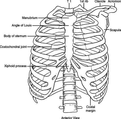
Both muscles attach to various ribs and parts of the spine. Roughly speaking, this is the area of the chest. Each true rib connects to its own strip of costal cartilage, which in turn connects to the sternum. Represents the anatomy of the ribs and muscle attachments. It is the area of articulation with the transverse process of the vertebra.
Pertinent Surgical Anatomy Of The Thorax And Mediastinum Anesthesia Key from aneskey.com Major landmarks of a typical rib are the following: Ten of the twelve ribs connect to strips of hyaline cartilage on the anterior side of the body. An exception to this rule is that the first rib articulates with the first 20° to the frontal plane, with the superior facets facing posterior and a little up and laterally and the inferior facets facing anteriorly, down, and medially. The thoracic cavity is made up of 12 pairs of ribs that connect in the posterior thorax to the vertebral bodies of the spinal column. Skeletal system anatomy and physiology nurseslabs. The posterior abdominal wall is a musculoskeletal structure formed by the posterior abdominal muscles, their fascia, the lumbar vertebrae and the image: The posterior end is composed of head, neck, and tubercle. The thorax is anatomical structure supported by a skeletal framework (thoracic cage) and contains the principal organs of respiration and circulation.
Further details of its anatomical relations and muscle attachments can be found in its own section in this text.
1.3 ribs anatomy and somatic dysfunctions. Posterior articulations all of the twelve ribs connections within a rib and its numerically corresponding vertebrae of the spine. The ribs form the main structure of the thoracic cage protecting the thoracic organs, however their main function is to aid respiration3. Test your knowledge about the ribs anatomy here Further details of its anatomical relations and muscle attachments can be found in its own section in this text. Gross anatomy there are 12 pairs of ribs which are separated by intercostal spaces. 12 pairs of ribs • 7 true ribs • 5 false ribs (including 2 floating ribs) •. Made up of thoracic vertebrae, ribs and… functions at upper end to connect the shoulder girdle and conn… Top suggestions for posterior ribs anatomy. They articulate with the vertebral column posteriorly, and terminate anteriorly as cartilage (known as costal cartilage). The posterior intercostal arteries anastomose with the anterior intercostal arteries to supply the structures of the thoracic wall. Its anterior ramus forms the intercostal nerves. The posterior abdominal wall is a musculoskeletal structure formed by the posterior abdominal muscles, their fascia, the lumbar vertebrae and the image:
They articulate with the vertebral column posteriorly, and terminate anteriorly as cartilage (known as costal cartilage). The lumbar plexus and its branches. by henry vandyke carter, henry gray (1918) anatomy of the human body. On the posterior side, your true ribs join with your thoracic vertebrae at the costovertebral and costotransverse joints. 12 pairs of ribs • 7 true ribs • 5 false ribs (including 2 floating ribs) •. The part of the muscle is thought to depress the ribs.
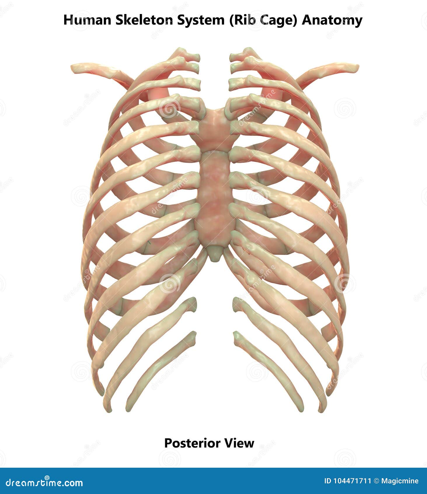
Human Skeleton System Rib Cage Posterior View Anatomy Stock Illustration Illustration Of Anatomical Health 104471711 from thumbs.dreamstime.com The thoracic cavity is made up of 12 pairs of ribs that connect in the posterior thorax to the vertebral bodies of the spinal column. Medial interchondral ligament of right seventh and eighth ribs. The rib below that is rib 2, and it connects to the t2 thoracic vertebra, and so on. The lower ribs are called floating ribs because they only attach to their corresponding vertebra on the posterior side; The ribs are a set of twelve paired bones which form the protective 'cage' of the thorax. In front, they are not attached, so they. They articulate with the vertebral column posteriorly, and terminate anteriorly as cartilage (known as costal cartilage). In vertebrate anatomy, ribs (latin:
1.3 ribs anatomy and somatic dysfunctions.
However, they do not attach directly to the sternum anteriorly, and instead, attach to the. The ribs are a set of twelve paired bones which form the protective 'cage' of the thorax. Gross anatomy there are 12 pairs of ribs which are separated by intercostal spaces. Ribs eight to ten are the false ribs and are connected to the sternum indirectly via the cartilage of the rib above them. Ribs 3 to 9 are considered typical ribs. Further details of its anatomical relations and muscle attachments can be found in its own section in this text. Its anterior ramus forms the intercostal nerves. The posterior end is composed of head, neck, and tubercle. Each true rib connects to its own strip of costal cartilage, which in turn connects to the sternum. The posterior cecal artery is located in the abdomen near the lower intestines. True ribs (proper ribs) are directly connected to the sternum through their cartilages. The subclavian artery and brachial plexus cross the rib posterior to anterior scalene muscle attachment and then run in contact with the bone on their way to the upper limb. In front, they are not attached, so they.
Be sure to subscribe to the visible body blog for more anatomy awesomeness! anatomy of ribs. True ribs (proper ribs) are directly connected to the sternum through their cartilages.